Digital X-ray

Less radiation, sharper images – for more precise diagnoses.

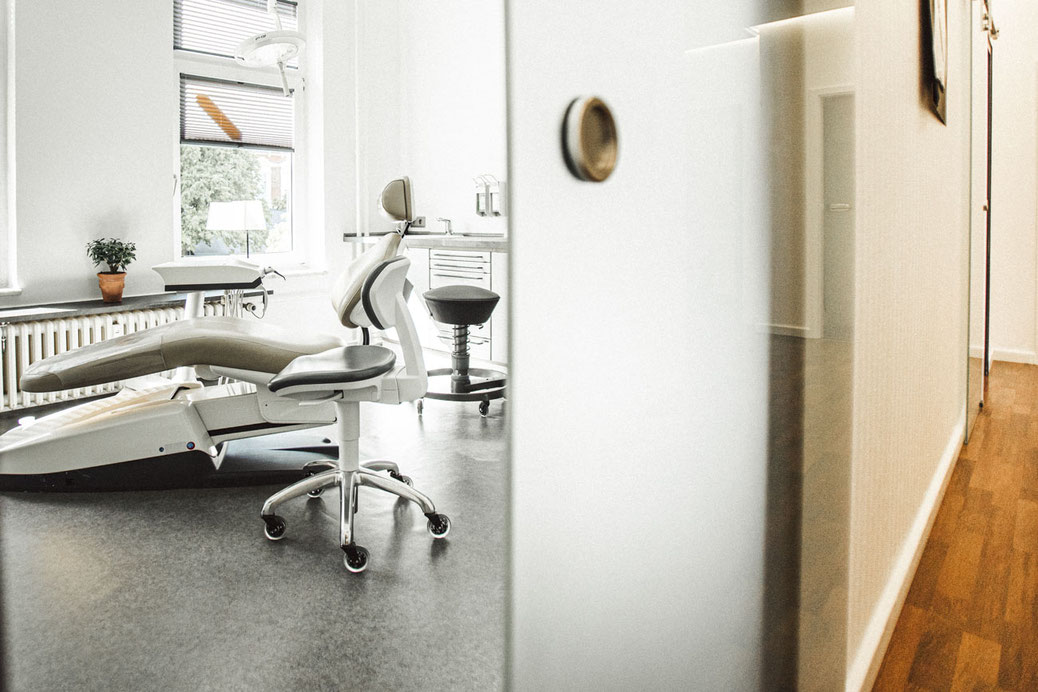
Digital X-ray system at the dental practice in Kiel

Digital X-ray

At our practice we work exclusively with digital X-ray technology. Compared with conventional X-ray film, it reduces radiation exposure for our patients by up to 90 percent – while producing noticeably sharper, more detailed images at the same time.

The digital images are available immediately on the treatment monitor. That means: no waiting time, no development chemicals, and an immediate discussion of the findings directly with you. We show you what we see – transparency is part of treatment for us.

  • Up to 90% less radiation exposure than conventional X-rays
  • High-resolution images for precise diagnosis
  • Instant image availability – no waiting, no development
  • Digital storage and easy comparison with previous images
  • Used for check-ups, fillings, implants and more
Book an appointment

When an X-ray makes sense

We don't X-ray routinely at every appointment, but specifically where an image actually improves diagnosis or treatment.

Decay diagnostics

Decay between the teeth is often not visible to the naked eye. A bitewing X-ray makes it visible before symptoms appear.

Root and bone findings

Before a root canal treatment or with unclear pain, the image shows the condition of the root, root tip and surrounding bone.

Monitoring periodontitis

Bone loss from periodontitis can be measured on the X-ray and compared over time – important for judging treatment success.

Planning tooth replacement

Before implants, crowns or bridges, the image shows bone volume and neighbouring structures – the basis for a precisely fitted plan.

Our principle: every X-ray is subject to a justified indication – we only X-ray when the diagnostic benefit clearly justifies the radiation exposure, and we document every image.

Frequently Asked Questions

Is digital X-ray really lower in radiation?
Yes. Digital X-ray sensors need significantly less radiation than classic X-ray film to produce an image that can be evaluated – at our practice up to 90% less than with conventional methods. In addition, every image is only taken when it's diagnostically necessary.
How often does X-raying need to happen?
That depends on individual findings and risk. There's no blanket rule of "once a year" – we decide case by case, for example on suspicion of decay between the teeth, before larger treatments, or when monitoring an ongoing therapy.
Is X-ray possible during pregnancy?
Dental X-rays cover only the head and neck area, far from the abdomen, and are only taken with a clear indication. Please tell us if you are pregnant anyway – we'll weigh together whether and when an image is necessary.
What does an X-ray show that an examination alone doesn't?
In a clinical examination we only see the tooth surface. Decay between the teeth, the condition of roots and bone, or hidden sources of inflammation only become visible on the X-ray. The two forms of examination complement each other.

State-of-the-art diagnostics for your peace of mind

Book an appointment – we'll examine and advise you with the latest technology.